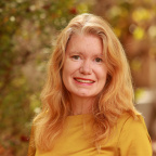
Speakers
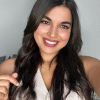
Solaima BELACHQER EL ATTAR
Technical University of Madrid
Visit Belachqer El Attar's Lab/Company Page
Bio: Dr. Solaima Belachqer-El Attar is an Assistant Professor at the Technical University of Madrid and a member of the GAqua Research Group. She holds an International Ph.D. in Biotechnology and Industrial Bioprocesses from the University of Almería, where her research on the solar chlorphoto-Fenton process was awarded the 2026 Best AOP Thesis Prize. Her work focuses on the scale-up of advanced oxidation processes for sustainable water reuse, resulting in the implementation of demonstration plants in real wastewater treatment facilities. Dr. Belachqer-El Attar has been recognized with the 1st Prize for Best Women Inventor by the Spanish Patent and Trademark Office (OEPM). A strong advocate for international collaboration, she has conducted research at Aalborg University and serves as deputy coordinator of the RII-AMAS network. She is the Principal Researcher of the SOL4REUSE project and an active contributor to European initiatives including LIFE PHOENIX and SFERA III projects.
Talk title: Belachqer-El Attar, S. et al. (2027). In progress.
Abstract: Every scientific career is, at its core, an ongoing experiment, one without a fixed plan, a guaranteed outcome, or a predetermined endpoint. This talk traces the trajectory of a young scientist who, in just five years, moved from a research fellowship to an assistant professorship, without ever losing sight of the question that started it all: how can we make water reuse a reality for rural water-stressed regions? The story follows the development of solar photo-Fenton technology from bench-scale reactors to demonstration plants implemented in real wastewater treatment facilities, a journey of scaling up not only photoreactors, but also confidence, collaborations, and scientific identity. However, this talk is not a list of achievements. It embraces the in progress, the failed experiments, the doubts between contracts, the teaching hours that shaped a vocation, and the "et al." of mentors, collaborators, and networks who made every milestone possible. Intended for early-career researchers, this offers not a blueprint but a reflection on building a life in research when the manuscript is not yet submitted and the best data may still be ahead.
Luiggi CAVALCANTI PESSÔA
SENAI CIMATEC University
Visit Cavalcanti Pessôa's Lab/Company Page
Bio: Luiggi Cavalcanti Pessôa is an Assistant Professor and Researcher at SENAI CIMATEC University (Brazil). His research focuses on circular bioeconomy, microalgae biotechnology, waste-to-value systems, biorefineries, and biogas and biomethane development. He holds an M.Sc. in Energy and Environment from Universidad Adolfo Ibáñez (Chile) and a Ph.D. in Chemical Engineering from the Federal University of Bahia (Brazil), including a visiting doctoral appointment at the University of Almería (Spain). Dr. Pessôa combines academic, industrial, and applied R&D experience, including leadership roles in manufacturing operations. He develops research grant proposals and leads collaborative research and innovation projects involving industry, government, and international partners, advancing resource recovery, circular economy, and energy transition solutions. He is the Technical Coordinator of the MBI in Circular Economy at SENAI CIMATEC and was recognized for his doctoral research with the 2024 UFBA Best Engineering Ph.D. Thesis Award and the 2025 UFBA Polytechnic School Recognition Award.
Title: From the Factory Floor to the Lecture Hall: An Unconventional Journey to Becoming a Young Professor
Abstract: More than once, I found myself standing on a factory floor, in different countries, convinced that industry would define my professional future. Yet one thing never went away: a persistent curiosity to keep growing, challenge myself, and better understand the world around me, even when I could not fully explain why. What followed was anything but a linear career path. A planned Ph.D. in Canada never materialized after scholarship funding was cut. I left industry to start over in a country whose language I did not speak. Personal losses and the passing of mentors forced me to rethink my priorities and sense of purpose. And when I finally secured a faculty position, a new international opportunity emerged, tempting me to change directions once again. This is not a talk about career planning. It is a story about uncertainty, curiosity, perhaps resilience, and how some of the most important decisions in life are often the ones we never intended to make. There was no roadmap. Looking back, many of the best decisions of my career were never part of the original plan. If these stories spark curiosity in even one person in the audience, then the journey will have been worthwhile. Hope you enjoy it!
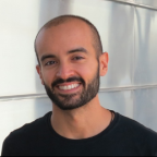
Isaac CANALS
University of Zurich – Kinderspital Zürich
Visit Canals' Lab/Company Page
Bio: I studied biology at University of Barcelona and received my PhD in Genetics in 2015 by the University of Barcelona working on Sanfilippo syndrome. During my postdoctoral training at Lund University, I specialized in neuroscience, stem cells and genome editing for modeling neurodegenerative disorders. I established my own research group in 2021 at Lund University and later at the University Children’s Hospital of Zurich in 2023. The aim of my research is to generate different iPSC in vitro models to understand disease mechanisms of lysosomal storage disorders, the contribution of different brain cells to the pathology and what are the neurodevelopmental impairments in patients, as well as to develop new therapeutic approaches based on genome editing technologies.
Talk title: Reprogramming Myself Along the Way
Abstract: In my talk I would give some insights into my journey from a bachelor student at University of Barcelona until the moment I was appointed Assistant Professor at University of Zurich, a journey through my PhD in Genetics at Barcelona and my postdoc and initial independence at Lund University. I will give my view on what steps worked well for me, what I could have done differently and how my experiences and interactions with more experienced researchers shaped my interests but also how I perform my research and my supervision now. A personal reprogramming journey within academia.
Felix RANDOW
MRC Laboratory of Molecular Biology, Cambridge, UK
Visit Randow's Lab/Company Page
Ubiquitylation of non-protein substrates: Mechanisms and consequences
Bio: Prof. Felix Randow is Professor of Molecular Immunity at the University of Cambridge and Programme Leader at the MRC Laboratory of Molecular Biology, where he leads a research programme investigating cell-autonomous and innate immunity. His research focuses on how host cells detect and eliminate intracellular bacterial pathogens, with particular emphasis on ubiquitin signalling, antibacterial autophagy, membrane damage sensing, and host–pathogen interactions. Over the past two decades, Prof. Randow has made several landmark discoveries that have transformed our understanding of innate immune defence mechanisms, including the identification of key pathways linking ubiquitin signalling and selective autophagy to antibacterial immunity. He is an elected EMBO Member and Fellow of the Academy of Medical Sciences, and has received numerous prestigious awards, including a Wellcome Trust Senior Investigator Award. His work has been published in leading journals such as Nature, Science, Cell, Nature Immunology and Molecular Cell.
Talk title: Ubiquitylation of non-protein substrates: Mechanisms and consequences
Abstract: Ubiquitylation is essential for eukaryotic life. Historically, proteins were thought to be the exclusive targets of ubiquitylation. Our discovery of lipopolysaccharide ubiquitylation during bacterial infection revealed non-protein ubiquitylation as an 'eat-me' signal for selective autophagy. In searching for endogenous substrates of non-protein ubiquitylation, we recently discovered the ubiquitylation of aberrant glycogen as an essential mechanism of glycogen quality control that prevents polyglucosan disease. I will discuss the mechanisms and biological significance of non-protein ubiquitylation in immunity and organismal homeostasis.
Astrid STUBBUSCH
Monash University, Clayton, Australia
Visit Stubbusch's Lab/Company Page
Prix Schläfli Award Ceremony and Lecture
Bio: Dr. Astrid Stubbusch is a microbial systems ecologist whose research explores how microbial metabolic strategies shape Earth’s biogeochemistry and climate. She is particularly fascinated by how microscopic processes scale up to drive ecosystem functioning and planetary-scale phenomena.
Astrid studied Biological Sciences (B.Sc.) and Systems Biology (M.Sc.) at Heidelberg University in Germany. During her studies, she had the opportunity to study and work abroad, first as an exchange student at the University of Leicester (UK), then as a research assistant at the University of Cambridge (UK), and later as a research assistant in Singapore. She completed her PhD at ETH Zürich and Eawag, the Swiss Federal Institute of Aquatic Science and Technology, in Switzerland. Her doctoral research focused on trophic interactions and nutrient acquisition strategies of bacteria.
To further investigate microbial metabolism and its significance for ecosystems, Astrid joined the Climate & Microbiology Group at Monash University in Melbourne as a postdoctoral researcher. There, she studied microbial hydrogen metabolism in the human gut and its role in health and disease. Supported by an SNSF Postdoc.Mobility Fellowship, Astrid currently explores how metabolic versatility influences microbial habitat ranges and shapes microbial responses to environmental changes.
Talk title: Nutrient Foraging Strategies in Microbial Communities
Abstract: Microorganisms inhabit environments where growth is frequently constrained by the availability of essential nutrients. To survive and thrive under these conditions, they have evolved diverse strategies to acquire and recycle resources, ranging from metabolic cooperation to competition and antagonism. Understanding these strategies is fundamental to explaining how microbial communities function and how they influence the cycling of matter and energy across ecosystems. Here, we will discuss recent insights into nutrient foraging in microbial communities, focusing on the discovery that bacteria can acquire nutrients by lysing neighboring cells through contact-dependent antagonism. Combining single-cell imaging, isotopic tracing, and ecological theory, this work reveals how microbial interactions can facilitate nutrient transfer between community members and support growth under nutrient-limited conditions. The findings suggest that antagonistic interactions may represent an important and widespread strategy for resource acquisition in natural microbial ecosystems. By placing these findings in a broader ecological context, the talk will explore how microbial nutrient foraging strategies shape carbon and nutrient fluxes across ecosystems, from local microbial communities to global biogeochemical cycles.
Georg Erich WINTER
AITHYRA – Research Institute for Biomedical Artificial Intelligence
Visit Winter's Lab/Company Page
Chemical Rewiring of Biological Circuits for Cancer Therapy
Bio: Georg Winter performed his graduate studies at CeMM in Vienna, working on elucidating the mechanism of action of cancer drugs with a specific emphasis on proteomics- as well as chemical genetics approaches. He continued his training in chemical biology, working as a postdoctoral fellow with Dr. James Bradner the Dana Farber Cancer Institute where he published the first paper reporting on in vivo target protein degradation and co-developed degron-tagging approaches that leverage the E3 ligase CRBN (dTAG approach) to understand the mechanistic involvement of gene control factors in oncogenic transcriptional circuits. He was recruited as a CeMM Principal Investigator in June 2016, continuing his work on targeted protein degradation with a particular emphasis on the phenotypic identification and mechanistic characterization of molecular glue degraders. In April 2025, Georg has been appointed as the Life Science Director at AITHYRA. The Winter lab has been funded via several national and international third-party grants, including two ERC Grants (Starting and Consolidator), an Aspire Award (Mark Foundation) and a Cancer Grand Challenge Award. Georg has authored more than 70 peer-reviewed papers and his contributions to the field of chemical biology in general, as well as targeted protein in particular, have been recognized via several international awards including the Eppendorf Award for Young European Investigators, the Wilson S. Stone Memorial Award from the MD Anderson, the Tetrahedron Young Investigator Award and the EFMC Price for Young Chemical Biologists.
Talk title: Chemical Rewiring of Biological Circuits for Cancer Therapy
Abstract: Cancer therapies have traditionally focused on inhibiting disease-driving proteins. An emerging alternative is to reprogram cellular circuitry by chemically rewiring protein function, interactions, localization, and fate. In this lecture, I will discuss recent advances in induced-proximity pharmacology and chemical biology that enable the rational manipulation of biological networks beyond conventional inhibition. Drawing on examples from targeted protein degradation, molecular glue discovery, transcriptional reprogramming, and chemically induced post-translational modifications, I will illustrate how small molecules can redirect endogenous cellular machinery to create new therapeutic mechanisms. These studies reveal opportunities to convert previously undruggable cancer dependencies into tractable therapeutic targets and suggest a broader framework in which medicines act not merely as inhibitors, but as programmable agents that rewire biological circuits. Together, these approaches point towards the possibility of re-imagining cancer therapy.
Magdalena GÖTZ
Institute of Stem Cell Research, Helmholtz Zentrum München and Physiological Genomics, Biomedical Center, Ludwig Maximilian University of Munich
Novel mechanisms of neurogenesis and neural repair
Bio: Prof. Dr. Magdalena Götz studied Biology and did her PhD at the FMI in Tübingen on the mechanisms of how input connections to the cerebral cortex form during development and how specific neuronal subtypes are specified. She then moved to the National Institute for Medical Research to use retroviral vectors for clonal analysis and identified mechanisms delineating neighboring forebrain regions. Afterwards she started her own lab at the Max-Planck Institute for Neurobiology and made the breakthrough discovery that radial glial cells are neural stem cells. This inspired her to attempt turning also mature glial cells into neurons already in 2002 in vitro and in 2005 in vivo. In order to determine which glial cells best to convert to neurons after traumatic brain injury, she systematically examined the roles of distinct glial subtypes after traumatic brain injury when she was appointed Director of the Institute of Stem Cell Research at the Helmholtz Center Munich in 2004 and Chair of Physiological Genomics, now at the Biomedical Center of the Ludwig-Maximilians University in Martinsried Munich. Since then she identified key hurdles in the glia-to-neuron reprogramming reprogramming which led to high efficiency in vivo reactive glia-to-neuron conversion as well as human glia-to-neuron conversion in vitro. As one of the hurdles she identified was metabolic conversion, this led her to identify the new concept of organelle heterogeneity in different cell types – from mitochondria to even centrosomes. This is exciting because protein localization at a specific organelle can explain how mutation of ubiquitous proteins can cause an organ-specific disease when mutated. Thus, she combines deep cell biological analysis with fate conversion mechanisms aiming to repair.
Talk title: Novel mechanisms of neurogenesis and neural repair
Abstract: Organelles such as centrosomes, nucleoli or mitochondria perform well-known common functions in all cell types. However, we found a surprisingly large degree of differences in composition in the context of development, disease and direct reprogramming. For example, the centrosome of human neural stem cells differs by more than half of its proteome from the one in neurons (O’Neill et al., Science 2022). Such cell type-specific composition also explains why some ubiquitous proteins have organ-specific defects, when mutated in patients, as they are only at a specific organelle in specific cell types. I will explain this for the splicing protein PRPF6 that plays specific roles at the centrosome in neural stem cells which is relevant for the disease periventricular heterotopia. I will then turn to organellar heterogeneity in direct neuronal reprogramming and discuss the role of mitochondria heterogeneity in this process and how to overcome hurdles in this conversion process due to late change of the mitochondrial proteome to a neuronal identity. I will then talk briefly about nucleolar heterogeneity, before turning to the cytoskeleton in the nucleus. I will talk about the discovery of a large number of microtubule-associated proteins in the nucleus, and discuss how the nuclear role of one of them affects neural stem cell fate and causes disease when mutated in patients. Taken together, these data highlight the concept of multiplying protein function with different roles at different organelles or cell compartments with relevance in development, disease and repair.
Alex M. JAEGER
Moffitt Cancer Center
Visit Jaeger's Lab/Company Page
Parallel Symposia Session I – From Proteome to Mechanism: In Vivo Insights into Cellular Biology
Bio: As an undergraduate at Ohio State University and graduate student at Duke University, my research focused on understanding protein function at a molecular level with biophysical techniques. During my postdoc at MIT, I uncovered an unexpected relationship between protein folding and MHC antigen presentation. As a Damon Runyon Postdoctoral Fellow in Tyler Jacks’ Lab, I integrated my protein biochemistry background with mouse model engineering and developed tools for lineage restricted analysis of antigen presentation in vivo. My independent lab has continued to expand our immunopeptidomics toolkit and have applied our unique approach to isolate MHC-I and MHC-II peptides from numerous in vivo contexts including healthy tissues and preclinical models of lung cancer, pancreas cancer, head and neck cancer, colorectal cancer, and melanoma. These efforts have yielded insights not only into the mechanisms that shape the antigen landscape of cancer but also identified therapeutically actionable targets for antigen-specific immunotherapy. Additionally, we have applied our MHC-I and MHC-II toolkit to isolate antigens from professional and non-professional antigen presenting cells including dendritic cells, macrophages, B cells, and fibroblasts. Finally, we have leveraged our interactions with clinical colleagues at Moffitt Cancer Center to analyze patient samples. These cross-species, integrated approaches have provided a holistic view of antigen presentation and revealed cell type specific programs of antigen processing and presentation that can be used to engineer antigen specific immunotherapies in cancer.
Talk title: Decoding antigen presentation in the tumor microenvironment
Abstract: Adaptive immunity to cancer requires effective antigen presentation on Major Histocompatibility Complexes (MHC). While recent advances in computational prediction of cancer-specific MHC antigens have ushered in a new era of antigen-specific immunotherapies, including cancer vaccines, there is widespread recognition that these approaches misidentify most actionable antigenic targets. To address this, many groups have employed liquid chromatography-tandem mass spectrometry (LC-MS/MS) based empirical measurement of peptides presented by MHC-I and MHC-II, also known as “immunopeptidomics”. These efforts have greatly expanded our understanding of antigen presentation across various cells and tissues. However, these analyses are typically performed with cultured cells or bulk tissues, limiting our understanding of cell-type specific patterns of antigen presentation in the tumor microenvironment (TME). Our group has pioneered a new approach, based on engineered Cre-inducible affinity-tagged mouse MHC-I or MHC-II (H2-K1, “KbStrep”; H2-Ab1, “AbStrep”), enabling lineage-restricted immunopeptidomics in vivo. We applied these tools to immunocompetent models of lung adenocarcinoma (LUAD) and pancreatic ductal adenocarcinoma (PDAC), enabling precise purification of MHC peptides from either malignant cells or various stromal cell lineages in the TME. Integrating immunopeptidomics data with single cell RNA sequencing facilitated the deconvolution of the cellular origin of various tumor associated antigens. Also, we applied next generation proteomics search strategies to capture highly complex landscapes of post-translationally modified MHC peptides, revealing additional peptide binding modes and reflecting intracellular oncogenic signaling throughout tumor evolution. Our data suggests that the antigen landscape is dependent on physiological context and is highly dynamic throughout tumor evolution, highlighting an ever-evolving landscape of MHC targets for T cells that may underlie the success or failure of immunotherapy. Understanding these patterns across cell types in the TME will likely yield more precise and effective antigen-specific immunotherapies. Beyond cancer, the application of lineage-restricted immunopeptidomics to models of infectious disease and autoimmunity has great potential for basic immunology discovery.
Celia BERKERS
Utrecht University, Faculty of Veterinary Medicine
Visit Berkers' Lab/Company Page
Parallel Symposia Session I – From Proteome to Mechanism: In Vivo Insights into Cellular Biology
Bio: Celia Berkers is Full Professor of Metabolomics and Vice Dean of Research at Utrecht University’s Faculty of Veterinary Medicine. Research in her team focusses on understanding metabolic re-wiring in the tumour microenvironment. Her aim is to leverage the power of metabolomics to identify novel metabolic targets with potential for exploitation by novel therapies. She is an expert in isotope tracing metabolomics (also known as fluxomics) and cross-omics data analysis, which she applies on complex in vitro model systems such as organoids and co-culture models, that her team develops for this purpose. Celia Berkers studied chemistry at Utrecht University, where she graduated with honours in 2003. She started her PhD at the Harvard Medical School in Boston, continued her research at the Netherlands Cancer Institute in Amsterdam, and received her PhD degree with honors in 2010. She then moved to the Beatson Institute for Cancer Research in Glasgow, where she did a post-doc in the laboratory of Prof. Karen Vousden. In 2013, Dr Berkers joined the Biomolecular Mass Spectrometry and Proteomics Department at Utrecht University as an independent group leader. She was appointed full professor at the Faculties of Veterinary Medicine and Science in 2018. She was awarded with the Antoni van Leeuwenhoek Prize, the Heineken Young Scientists Award for Biochemistry and Biophysics, and was elected member of the Young Academy of the Royal Society. She is an Associate Editor for Cancer & Metabolism and an Editorial Board Member for Molecular Omics.
Talk title: tba
Abstract: tba
Carolin LERCHENMÜLLER
Chair for Gender Medicine, University of Zürich and University Hospital Zürich
Visit Lerchenmüller's Lab/Company Page
Parallel Symposia Session I – Gender Health in Biomedical Research: Advancing Precision Therapeutics
Bio: Carolin Lerchenmüller is Professor and Chair of Gender Medicine at the University of Zurich and a cardiologist at the University Hospital Zurich, where she leads the Women’s Heart Health Program within the Department of Cardiology.Following her medical studies and specialist training in Heidelberg, she conducted research at Massachusetts General Hospital and Harvard Medical School. Her research combines molecular cardiology and gender medicine, focusing on sex and gender differences in cardiac adaptation, disease, and injury, as well as cardioprotective mechanisms of the heart.In addition, her work addresses the role of gender medicine in global health, as well as issues of equity, diversity, and inclusion in medicine and science.
Talk title: From Mechanisms to Medicines: Integrating Sex and Gender in Biomedical Research
Abstract: Sex- and gender-sensitive medicine recognizes biological sex and sociocultural gender as distinct but interacting determinants of health and disease. It examines how sex-related biology influences molecular pathways, disease mechanisms, and responses to drugs, while gender shapes exposures, health behaviours, access to care, and treatment.Integrating these dimensions across the research continuum - from cells and animal models to clinical trials and clinical practice - is essential for reproducible science and more precise, effective therapeutics. In this talk, I will provide an overview of the current state of the field and discuss how sex and gender can be appropriately incorporated at different stages of biomedical discovery. Drawing on our research in cardiovascular medicine, clinical trials, and the distribution of research efforts, I will illustrate how sex- and gender-informed approaches can reveal biological mechanisms, identify persistent blind spots, and improve the translation of biomedical research into better care for everyone.
Barbora VIDIMOVA
University of Lausanne
Visit Vidimova's Lab/Company Page
Bio: I am a PhD student in Quantitative Biology at the University of Lausanne. My doctoral research investigates dendritic spine pathology in intellectual disabilities, with a focus on developing deep learning tools for high-resolution microscopy image analysis. A key aspect of my work is the implementation of the 3R principles by replacing mouse models with human organoids and establishing reproducible analytical pipelines for quantitative imaging.
Talk title: Self-supervised deep learning reveals novel physiological and pathological states of dendritic spines
Abstract: Dendritic spine morphology and motility, quantified from time-resolved microscopy, provide key readouts of diverse physiological and pathological states. Yet, existing image-analysis pipelines remain dependent on manual spine detection and classification into predefined categories, failing to capture spines’ full phenotypic diversity and limiting analytical reproducibility. Here, we introduce 4D MORPHS, a deep-learning framework designed to provide automated, scalable, and unbiased analysis of dendritic spine structure and remodelling in live microscopy images.4D MORPHS combines deep-learning-based segmentation and tracking with a self-supervised vision transformer that represents individual spines as latent embeddings, enabling data-driven identification of spine phenotypes without predefined classes. Across experimental datasets, the framework identified 12 distinct spine states and quantified their structural transitions over time. When applied to mouse neurons and human brain organoid models of fragile X syndrome, 4D MORPHS revealed disease-associated, previously unrecognized spine phenotypes. The framework also detected chemically induced alterations in spine morphodynamics. By enabling sensitive, standardized phenotyping at single-spine resolution, 4D MORPHS maximises the information obtained from each biological sample and reduces reliance on manual and potentially variable analysis. This approach provides a reproducible platform for evaluating human-relevant models of synaptic dysfunction and for identifying candidate structural biomarkers while supporting more efficient use of experimental animals.
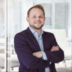
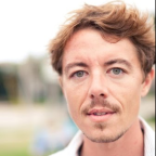
Martin PACESA
University of Zurich
Visit Pacesa's Lab/Company Page
Bio: My group’s focus is on investigating the dynamics of biomolecular assemblies and trying to design them from the ground up using computational protein design.
Talk title: Accurate design of protein binders
Abstract: Protein-protein interactions are central to biology and drug discovery, yet traditional antibody generation is slow and costly. BindCraft is an open-source, automated computational pipeline for de novo protein binder design that routinely yields nanomolar binders with 10-100% experimental success, without high-throughput screening or maturation. We illustrate their different biological and biochemical applications and outline how deep learning workflows can accelerate next-generation therapeutics, diagnostics, and bioprocessing.
Ludovic MURE
Inselspital and University of Bern
Parallel Symposia Session II – Neuronal Circuit Vulnerability Underlying Neurodegeneration
Bio: Ludovic Mure completed his MS in Neuroscience at the University of Lyon, France, and his Ph.D. in the laboratory of Dr. Howard Cooper at the SRBI (INSERM) where he investigated the recently discovered melanopsin, a photopigment expressed by a subset of retinal ganglion cells and rendering them intrinsically photosensitive (ipRGCs). He pursued in this very exciting and fast-moving field by joining the lab of Satchin Panda (The Salk Institute, San Diego, US). As a postdoc, he explored the non-visual responses to light in particular the relationship between the circadian clock and light. He found that melanopsin unique properties relied on both its structure and its binding partners. Then, in an exceptional collaborative project between INSERM, the Salk Institute, and the Institute of Primate Research (Kenya), he established the first rhythmic transcriptome of a diurnal primate. This study unveiled the unique features of primate rhythmic gene expression and unveiled the importance of the natural light-dark cycles for gene expression. Recently, together with F. Vinberg (U. of Utah) and A. Hanneken (Scripps Institute), he developed a human retina preparation that preserves the function of each photoreceptive system for several hours after the death of the donors and was able to record for the first time human ipRGCs and to characterize their responses, sensitivity, spectra, and functional diversity. Finally, in 2021, to transition to an independent research position in Europe, he joined the labs of Profs. Kleinlogel (U. of Bern) and Adamantidis (Department for Neurology, Inselspital) as a Velux Stiftung fellow to dissect ipRGCs circuits in the retina and the brain, in health and disease. In 2023, he joined the Department of Ophthalmology of the Inselspital in Bern (Switzerland) as a group leader.
Talk title: Alzheimer’s Disease is casting a shadow over the brain
Abstract: Sleep and circadian disruptions are among the earliest manifestations of Alzheimer’s disease (AD) and may emerge years before cognitive symptoms. Circadian rhythms allow physiology and behavior to adapt to the day–night cycle. Light is the main environmental signal that synchronizes the circadian clock to the 24-hour day and is detected by a specialized class of intrinsically photosensitive retinal ganglion cells (ipRGCs). Recent studies have revealed a selective loss of ipRGCs and alterations in their morphology in patients with AD. At the same time, light interventions can improve sleep and circadian outcomes in patients with AD. We therefore hypothesized that retinal light signaling to the brain through ipRGCs is progressively altered in AD. We combine histopathological analyses and functional recordings from human donor retinas and a mouse model of AD to examine ipRGC structure and function across disease progression. We find a reduced number of ipRGCs in AD, together with marked morphological alterations in surviving cells. Importantly, these surviving ipRGCs also display abnormal responses to light, revealing functional impairment of this pathway in addition to structural degeneration. Alterations in ipRGCs and their circuits may contribute to the sleep and circadian disturbances observed in patients with AD. Characterizing ipRGC-dependent responses to light may therefore provide new insight into the early pathophysiology of AD, identify accessible functional markers of disease progression, and ultimately help inform evidence-based light interventions aimed at preserving sleep and circadian function during healthy and pathological aging.
Sabine LIEBSCHER
Institute of Systems Neuroscience, Medical University Innsbruck, Innsbruck, Austria
Visit Liebscher's Lab/Company Page
Parallel Symposia Session II – Neuronal Circuit Vulnerability Underlying Neurodegeneration
Bio: Sabine Liebscher is a physician and neuroscientist, currently Professor of Systems Neurobiology and Director of the Institute of Systems Neuroscience at the Medical University of Innsbruck, a position she has held since February 2025. Before her appointment in Innsbruck, she served as W2 Professor of Cellular Neurophysiology at the Faculty of Medicine, University of Cologne, and previously led a Clinician Scientist research group while training in Neurology at the Institute of Clinical Neuroimmunology, at the University hospital Munich of the Ludwig Maximilians University (LMU) Munich, Germany. She studied medicine at the Technical University of Dresden, where she also completed her MD thesis. In 2007, she joined the laboratory of Nobel Laureate Paul Greengard at The Rockefeller University in New York. She later earned her PhD in Neuroscience with distinction from the Max Planck Institute of Neurobiology and the LMU Munich. In 2014, she established her independent junior research group through the prestigious Emmy Noether Programme of the German Research Foundation (DFG). Her research focuses on the pathophysiology of neurodegenerative diseases, particularly amyotrophic lateral sclerosis (ALS). She investigates how neural circuits and their components contribute to disease symptoms and drive the degenerative process. Her work integrates clinical insight with experimental neuroscience, combining in vivo two-photon imaging, single-cell transcriptomics, and viral circuit manipulation to uncover network-level mechanisms of neurodegeneration.
Talk title: Exciting complexity: Circuit mechanisms of cortical dysfunction in ALS / FTD
Abstract: Neurodegenerative diseases are increasingly recognized as disorders of neural circuits rather than the simple consequence of cell loss. Functional alterations in defined neuronal populations and networks often emerge before overt degeneration and can persist beyond it. Our work focuses on identifying these circuit-level dysfunctions to uncover early pathogenic mechanisms and therapeutic targets. I will present examples from amyotrophic lateral sclerosis (ALS), the most common adult-onset motor neuron disease, characterized by the loss of upper and lower motor neurons. A hallmark of ALS is cortical hyperexcitability, which appears early and plays a pivotal role in disease progression. Our studies show that this hyperexcitability is associated with early synaptic alterations and hyperresponsive layer 2/3 pyramidal neurons, which provide excessive glutamatergic drive to upper motor neurons and thereby promote aberrant feedforward excitation. In parallel, we find that noradrenergic input to the motor cortex is markedly reduced in both mouse models and ALS patients, suggesting a loss of neuromodulatory control that may further exacerbate cortical hyperexcitability. Together, these alterations arise before overt neuronal loss and contribute to abnormal motor output, indicating that network dysfunction, rather than degeneration alone, underlies selective vulnerability. Targeting compromised circuit elements delays disease onset and progression, underscoring their pathophysiological importance.
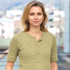
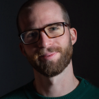
Lucas PAOLI
School of Life Sciences, EPFL
Visit Paoli's Lab/Company Page
Parallel Symposia Session II – Self-assembly Across Scales: from Cells to Ecosystems
Bio: Lucas Paoli is an ELISIR group leader at the School of Life Sciences of EPFL. His lab uses environmental genomics to study the diversity and ecology of microbiomes. Through large-scale, data-driven approaches, they aim to understand how ecology shapes microbial immunity across environments. Prior to that, Lucas was an EMBO Postdoctoral Fellow with Dr. Aude Bernheim at Institut Pasteur in Paris, where he worked on microbial immunity. He holds a MSc in Ecology and Evolution, a MPhil in Environmental Policy, and conducted his PhD at ETH Zürich under the guidance of Prof. Dr. Shinichi Sunagawa. His doctoral work helped decipher the genomic blueprint and biosynthetic potential of the global ocean microbiome.
Talk title: Ecological diversity of microbial immunity
Abstract: Bacteria and archaea have evolved numerous defense pathways to fend off infections by viruses and other genetic elements. However, the current census of prokaryotic immune diversity is heavily biased towards cultivated lineages, focuses on molecular mechanisms or evolutionary conservation, and largely blind to ecological distribution. Here, we screened 3.9 million genomes, including >2 million recovered from over 10,000 metagenomic samples, and identified 17 million defense systems. We then propose a framework to organise this diversity in defense system families using unsupervised iterative clustering at scale. With this approach, we identify 300,000 defense system families across microbiomes, find distinct immune repertoires across ecosystems, and track immune dynamics in the human gut. This works contributes to a new picture of prokaryotic immunity by providing an ecological lens at global scale.
Andreas DENDORFER
Ludwig-Maximilians-University München, LMU Hospital, Walter-Brendel-Centre of Experimental medicine
Visit Dendorfer's Lab/Company Page
Bio: Andreas Dendorfer is Professor for Cellular Signal Transduction at the Walter-Brendel-Centre of the LMU Hospital. He studied electrical engineering and medicine, and received his professional education at the Institute of Physiology of the LMU München, and the Institute of Pharmacology and Toxicology of the Medical University of Lübeck. His research interests are focused on cardiovascular physiology and pharmacology, covering hypertension, autonomic innervation, and cardiac metabolism and preconditioning. More recently, he established the use of myocardial living slices for his research, and developed a bioreactor for the biomimetic cultivation of this tissue preparation. To promote tissue culture technology in general, he co-founded the start-up company InVitroSys.
Talk title: Biomimetic culture of human myocardium: A versatile in vitro model
Abstract: Living myocardial slices (LMS) are thin, vibratome-cut sections of authentic myocardium which can be prepared from hearts of various species, and particularly, from explanted human myocardium of heart transplant recipients. Since O2 demands are covered by diffusion, the viability and functional state of LMS can be maintained under biomimetic conditions (preload, pacing, compliance) for many weeks. As such, LMS form a medium complexity experimental platform which provides a 3-dimensional, multicellular, biomechanical model of adult myocardium, but excludes exogeneous factors such as perfusion, metabolism, and neuro-hormonal regulation. The experimental benefits of the model comprise long-term interventions, sequential functional readouts, and free end-point analyses.LMS have been used for studies on drug effects, cardiac plasticity, cellular interactions, and genetic cardiomyopathies. They exclusively enable such investigations in human adult myocardium. However, the model presents limitations due to the low availability and pathologic state of human myocardial samples, and to the artificial conditions of tissue culture. The vision to meet these challenges includes strict definition of suitable study protocols, highly specific interventions, and high content analyses. Perspectively, a standardized experimental and analytical workflow needs to be established in order to develop the full potential of the cardiac slice culture in between cellular models and in vivo experiments.
Olivier GUENAT
University of Bern, ARTORG Center
Visit Guenat's Lab/Company Page
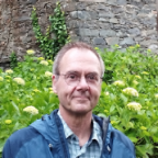
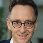
Bio: Olivier T. Guenat is Professor in Biomedical Engineering at the University of Bern in Switzerland and Head of the Organs-on-Chip Technologies Group at the ARTORG Center. He is associated with the Pulmonary Medicine and the Thoracic Surgery Divisions of the University Hospital of Bern. His research focuses on the development of organs-on-chip, in particular lung-on-chip models that mimic the healthy and diseased in-vivo cellular microenvironments of the lung parenchyma. Prior to his position at the University of Bern, he worked at the Swiss Centre for Electronics and Microelectronics (CSEM), was an assistant professor at Ecole Polytechnique de Montréal (QC, Canada), and did postdoctoral work at Harvard Medical School in Boston and at the University of Neuchâtel in Switzerland. He is the founder of AlveoliX and co-founder of Vitronco, two biotech start-ups that spun out of his lab.
Talk title: From capillaries to arterioles on chip: multicellular vascular platforms with functional read-outs
Abstract: We present a suite of multicellular vascular platforms capturing distinct aspects of human microcirculation. Self‑assembled microvessels formed by vasculogenesis generate perfusable capillary‑like networks with physiological endothelial barrier and transport functions. A second platform applies cyclic mechanical stress to these vessels, revealing strain‑dependent changes in permeability, alignment, and gene expression relevant to cardio‑pulmonary environments. Finally, preliminary results of an arteriole‑on‑chip with circumferentially aligned smooth muscle cells enabling controlled vasoactive responses will be presented. Together, these complementary systems provide functional vascular readouts across multiple scales of the human microvasculature.
Nadège ZANOU
University of Lausanne
Visit Zanou's Lab/Company Page
Bio: Dr. Zanou is an M.D., Ph.D., and Senior Lecturer - Group Leader at the University of Lausanne. She received her medical degree in Benin in 2006 and then completed master's and Ph.D training in biomedical sciences at the Catholic University of Louvain in Belgium, earning her Ph.D. in 2012. During her Ph.D, Dr Zanou's research focused on the role of plasma membrane calcium channels in skeletal muscle development and function, as well as in the pathophysiology of Duchenne muscular dystrophy. In 2013, she received a research associate fellowship from the Belgian National Fund for Scientific Research to continue her research on the activation mechanisms of those channels in normal and pathological cells. In 2015, her work received an award from the Académie Royale de Médecine de Belgique. She then moved to Switzerland to broaden her international experience. In Lausanne, Dr Zanou’s research focused on intracellular calcium channels, especially the role of the ryanodine receptor type 1 (RyR1) channels in muscle health and disease. She developed the Institute of Sport Sciences' first molecular laboratory at the University of Lausanne, enabling her to conduct translational research on muscle adaptations to exercise and disease and the role of intracellular calcium channels including the RyR1. She also completed research internships at the University of Barcelona in Spain (2017) and at Columbia University in the United States (2017 and 2018). Her success is reflected in her numerous high-impact publications and robust collaborations forged locally in Lausanne (UNIL, EPFL, Nestlé Research, and CHUV) and abroad. She received several competitive grants (Swiss National Science Foundation grant, UNIL-CHUV Interdisciplinary grant, Swiss Foundation for Research on Muscle Diseases grant…) to support her different research projects.
Talk title: The ryanodine receptor at the hub of skeletal muscle plasticity to exercise and disease
Abstract: The ryanodine receptor type 1 (RyR1) is the primary intracellular calcium release channel in skeletal muscle cells. It is located at the endoplasmic/sarcoplasmic reticulum (ER/SR) and is involved in excitation-contraction coupling (ECC). RyR1 is stabilized by a protein called calstabin1 (calcium channel stabilizing binding protein 1), which is encoded by FKBP12. RyR1 function can be modulated by post-translational modifications, such as oxidation, phosphorylation, or nitrosylation, which lead to calstabin1 dissociation and leaky RyR1. Alternatively, mutations can alter RyR1 configuration or expression. Prolonged leaky RyR1 has been shown to play a detrimental role in several diseases.Using a translational approach that combined experiments in human and mouse models, as well as in vitro models of simulated exercise, we showed that a single session of sprint interval training (SIT) triggers acute Ca²⁺ leak through RyR1, whereas moderate-intensity continuous training (MICT) does not. The mitochondria take up this Ca²⁺ to dephosphorylate and activate pyruvate dehydrogenase (PDH), thus enhancing NADH-linked mitochondrial bioenergetics.

Ivan MARTIN
Department of Biomedicine, University of Basel
Visit Martin's Lab/Company Page
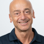
Bio: Prof. Dr. Ivan Martin studied Biomedical Engineering at the University of Genova where he obtained his PhD in 1996. Between 1996 and 1999 he was a postdoctoral associate at Harvard/MIT. He joined the Department of Biomedicine (DBM) at the University Hospital of Basel in 1999 as leader of the Tissue Engineering Research Group, in close coordination with the surgical units. In 2007 he was appointed Professor for Tissue Engineering at the University of Basel and from 2021 he is Director of the DBM. From 2004 to 2009 he was the first president of the European section of the Tissue Engineering Regenerative Medicine International Society (TERMIS), and later Chair of the TERMIS Strategic Alliance Committee. In 2018 he was elected as member of the Swiss Academy of Medical Sciences. He has been Chair of the ‘Mesenchymal stromal cell committee’ and is currently member of the ‘Orthopaedics and Musculoskeletal Committee’ of the International Society for Cellular and Gene Therapy (ISCT). He is part of the editorial boards of 6 international journals. His group includes scientists from the biological, engineering and clinical fields, dedicated to develop solid scientific basis for innovative translational strategies in regenerative surgery. In this field he is author of more than 300 peer-reviewed papers on international journals (H-index of 109), inventor on 12 patent applications and recipient of grants from the main funding bodies, including the prestigious European Research Council. The developed science and technology have been translated into different clinical trials for cell-based cartilage and bone repair, and into the founding of a spin-out company for the commercialization of bioreactors for 3D cell culture (Cellec Biotek AG).
Talk title: Bone and Bone Marrow Engineering
Abstract: The lecture will present the development and translational trajectory to robustly generate bone tissue through the recapitulation of endochondral ossification. The strategy, which implies the chondrogenic differentiation of mesenchymal stromal cells derived from human bone marrow or adipose tissue, has offered the opportunity not only to generate osteoinductive grafts, but also to address fundamental questions related to the process of limb development in a fully human system. In particular, insights have been generated on the onset and function of ‘skeletal stem cells’ in an environment mimicking the growth plate in developing bones. The lecture will also present a recently engineered iPS cells-derived 3D culture system capable to model the osteoblastic, endothelial and neural cellular elements of the bone marrow niche. The platform has been used to investigate how specific elements of the bone marrow niche regulate engraftment and lineage specification of hematopoietic stem/progenitor cells. The engineered model is currently being extended to investigate niche-mediated processes in specific malignancies, including chemoresistance.
David BRÜCKNER
Biozentrum & Department of Physics, University of Basel
Visit Brückner's Lab/Company Page
Parallel Symposia Session III – Mini-symposium: Decoding Multicellular Self-Organization
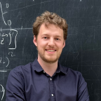
Bio: David Brückner is an Assistant Professor of Theoretical Biophysics at the Biozentrum and Department of Physics of the University of Basel, Switzerland. His research focusses on theoretical physics approaches to uncover how interactions of cells and molecules control the behavior of multicellular systems. Previously, he was Postdoc at the Institute of Science and Technology Austria and obtained his PhD from the Ludwig Maximilian University in Munich. He has been awarded with the Gustav-Hertz-Prize of the German Physical Society, and received an ERC Starting Grant.
Talk title: Information flow in self-organized developmental systems
Abstract: Embryonic development relies on multi-cellular systems self-organizing into precise spatial patterns of cell fates. An inevitable obstacle is intrinsic noise at the single-cell level, which constrains the information available to cells for fate decisions. This information is often considered to be encoded in extracellular morphogen concentrations, but cells respond to a much broader set of inputs, including dynamical and combinatorial signaling as well as mechanosensitive pathways that sense tissue mechanics. Yet we lack principled frameworks to quantify and predict how cells obtain sufficient information to reliably differentiate into the right fate at the right time and place. I will discuss how combining biochemical and mechanical models of patterning and morphogenesis with information theory provides a mathematical language for analyzing biological self-organization across diverse systems. First, I will demonstrate how the coupling between mechanosensitive YAP1 signaling and lateral inhibition governs diverse epithelial organoids, from intestinal to mammary. We develop a unifying model for these systems and analyze it using information theory. Second, I will discuss how richer inputs, from mechanical gradients to combinatorial signaling, can be incorporated into this framework. This opens an avenue toward unifying the zoo of chemical and mechanical signaling processes that orchestrate embryonic development.
Elda BAUDA
University of Geneva
Visit Bauda's Lab/Company Page
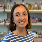
Bio: My research aims to investigate the cellular envelope of unicellular organisms such as bacteria and yeast and to understand how the constraints of unicellular life necessitate rapid and robust adaptation to environmental stress. Despite their importance as protective barriers and as dynamic regulatory platforms, the native architecture of cellular envelopes remains poorly understood. To address this, I use and further develop in situ structural cell biology approaches, especially cryo-FIB-ET, which has recently opened a new frontier in exploratory imaging. By integrating cryo-FIB-ET with molecular cloning and live fluorescence microscopy, my long-term goal is to establish a structural cell biology framework that links membrane architecture to cellular decision-making in bacteria and yeast.
Talk title: Thekosomes: a new cellular strategy for membrane tension control
Abstract: The plasma membrane (PM) is the primary interface through which cells sense and respond to environmental stress, making its homeostasis essential for survival. Over the years, PM invaginations in S. cerevisiae have been reported across diverse experimental settings and under different names, particularly in conditions of reduced membrane tension. However, these observations have remained fragmented, lacking a coherent interpretation. Here, we integrate these seemingly disparate findings by demonstrating that membrane invaginations arising under low PM tension define a single, previously unrecognized cellular compartment, which we term thekosomes. Using cryo-focused ion beam electron tomography (cryo-FIB-ET), we reveal that thekosomes are membrane-bound compartments spanning hundreds of nanometers. Their lumen, containing organelles and proteins, is enclosed from the cytoplasm by two membranes incorporating cell wall material. Their formation is rapid and relies only on biophysical property change of the membrane. We demonstrate the recruitment of key membrane-associated proteins, including TORC2, Slm1, and septins, to thekosome upon their formation. Under conditions of chronic disruption of PM homeostasis, thekosomes are constitutively present at the PM. We further reveal thekosomes in the distantly related S. pombe, underlying a conserved membrane stress response mechanism. Together, these findings reveal highly dynamic, organelle-containing compartments that couple membrane remodeling and lipid homeostasis to maintain cellular integrity under stress.
Amy GLADFELTER
Duke University, USA
Visit Gladfelter's Lab/Company Page
Bio: Amy Gladfelter is a quantitative cell biologist interested in fundamental mechanisms of cell organization. She is currently a Distinguished Duke Health Science and Technology Professor in the Cell Biology and Biomedical Engineering Departments at Duke University. Previously she was Professor of Biology and Associate chair at the University of North Carolina at Chapel Hill from 2016- 2023. She remains a longstanding fellow of the Marine Biological Laboratory in Woods Hole, MA and is currently the course director of the Physiology Course. In her research program, she uses microscopy, biophysical and genetic approaches to study syncytial cells and how these large cells spatially organize the cytoplasm via biomolecular condensates and sense their shape.
She has been honored with the 2014 Graduate Mentoring Award from Dartmouth, the 2015 Mid-Career Award for Excellence in Research from the American Society of Cell Biology, the 2020 Graduate school mentoring award from UNC and was a Howard Hughes Medical Institute Faculty Scholar. She is an elected fellow of AAAS, the America Academy of Microbiology and the American Academy for Arts and Sciences and member of the National Academy of Sciences.
Talk title: Cellular computing using condensates
Abstract: Syncytia are found throughout the biosphere from free-living fungi to the human placenta and malignant tumors. These cells contain many nuclei in a shared cytoplasm raising the possibility of genome cooperation and competition even within a shared cytoplasm. Work in our lab focuses on understanding the emergent functions that arise from having multiple genomes in the same cell in both filamentous fungi and in the human placenta. One area of work is focused on how the cytoplasm is structured to support different nuclear identities using condensed assemblies of RNA and proteins. Biomolecular condensates are vessels to spatially and temporally control biochemistry in cells for a variety of processes. Despite their ubiquitous roles in the entire lifecycle of RNA, how RNA impacts the composition, properties and functions of condensates remains poorly understood. The chemical properties and sequence of RNAs are generally considered in light of housing or regulating the genetic code. However, most amino acids are encoded by multiple codons, making the genetic code degenerate. Synonymous mutations in RNA sequences affect protein translation and folding, but their impact on RNA itself is often neglected. We developed a genetic algorithm that introduces synonymous mutations to control the conformational heterogeneity of structures sampled by an mRNA. The behavior of the designed mRNAs reveals physical information layered in the genetic code. We find that mRNA conformational heterogeneity impacts the physical properties and functional outputs of RNA-protein complexes and biomolecular condensates. The role of structure and disorder of proteins in biomolecular condensates is well appreciated, but we find that RNA conformational heterogeneity is a major contributor to condensate properties. This feature of RNA enables both evolution and engineers to build cellular structures with specific material and responsive properties. This talk will examine how syncytial cell organization leads to distinct kinds of cell functions and how RNA in condensates is used to spatially pattern functions in these giant cells.