Meeting Schedule
Program
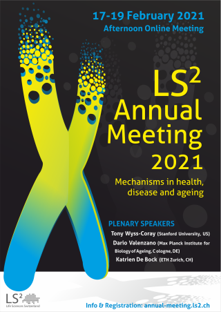
Please find below the schedule of the LS2 Annual e-Meeting 2021 (click on the image to download the program, scroll down this page for session details .
Details of the Scientific e-Symposia and abstracts of invited and industry speakers here
Details of the Young Scientists' e-Symposium here
Details of speakers here
List of e-posters here
Abstracts of plenary here
| Wednesday | 17.02.202113:00 – 18:30 |
| 13:00 – 18:30 | Wednesday's Program |
Welcome Words Welcome Words Mario Tschan (UNIBE) as chair of the LS2 Annual e-Meeting 2021 and Didier Picard as president of LS2 will open the meeting | |
Plenary Lecture I African killifishes shed new light on life history evolution and reveal an important role of the gut microbiota in modulating vertebrate lifespan Species in nature display a staggering diversity in individual lifespan. From mayfly that live a few hours after hatching, to thousands-year old plants that keep reproducing throughout their life. Lifespan in a given species is limited by external factors, such as predation, infections, starvation, dehydration, etc., as well as by internal factors, including molecular, cellular and tissue damage caused by natural biological processes, such as cell replication and accumulation of aberrant sub-cellular protein aggregates. In my presentation, I will discuss the case of African killifishes, a unique group of fishes that have repeatedly adapted their life cycle to survive in extreme environments, represented by savannah water pools that completely desiccate once a year during the dry season. Among African killifishes, the turquoise killifish (Nothobranchius furzeri) is the shortest-lived species, with a lifespan that ranges from four to eight months. I will describe the life cycle of the turquoise killifish and explain what it means for this fish to undergo “aging”. In the final part of my talk I will present our recent findings on how the gut microbiota plays a causal role in modulating aging and lifespan and how understanding the interaction between adaptive immune system and the microbiota gives us novel insights into the biology of aging and offers new possibilities for future anti-aging therapeutic interventions. | |
Plenary Lecture II. Prix Schläfli Award Alice BERHIN - WINNER PRIX SCHLÄFLI 2020 Louvain Institute of Biomolecular Science and Technology (Université Catholique de Louvain - BE) Visit Berhin - Winner Prix Schläfli 2020's Lab Page Discovery of the Root Cap Cuticle: Structure and Functions. During land colonization, plants developed extracellular diffusion barriers, such as cuticle to isolate themselves from the environment. The cuticle is a lipid layer, made of cutin, deposited at the surface of the plant shoot. It prevents the loss of water and nutrients and protect plants against stresses. Plant cuticles have been studied since the 19th century and only characterized in the shoot epidermal cells. A cuticle at the root was not imagined to be compatible with its uptake function. Hence, the specific localization of the cuticle at the epidermis of the aerial organ became a defined feature of the cuticle. However, the intriguing expression of genes involved in cutin biosynthesis at the tip of primary and lateral roots of Arabidopsis has led to the investigation of the cell wall ultrastructure at the root cap cells, the outer cell layer of the root tip. The root cap cells of young primary roots and lateral roots of Arabidopsis, as well as of other species, are covered by an layer highly similar to the Arabidopsis leaf cuticle. The structure, the composition and the biosynthesis pathway of the root cap cuticle was investigated. The root cap cuticle of young primary and emerging lateral roots plays important roles in root physiology and development such as diffusion barrier protecting the root meristem from toxic compounds and the reduction of organ adhesion causing a delay in lateral root emergence. Until now, plant cuticles of different aerial organs have been exclusively associated with epidermal tissues of the shoot, our discovery of a cuticle at the root cap now challenges this dogma and adds a new element to our understanding of root anatomy, development, and physiology. | |
Plenary Lecture III. Friedich - Miescher Award Andrea Ablasser Sensing DNA as a danger signal through the cGAS-STING pathway The life of any organism depends on the ability of its cells to recognize and respond to pathogenic microbes. To accomplish this vital task cells rely on intricate signaling pathways that couple sensing of pathogen-associated danger signals to the execution of antimicrobial immune responses. The cGAS-(cGAMP)-STING signaling pathway constitutes a highly conserved innate immune sensing strategy that originated in bacteria to protect from phage infection. In mammals, the pathway detects intracellular DNA to then initiate an antiviral and inflammatory state. It is becoming increasingly apparent that the cGAS-STING pathway plays a critical role in regulating a number of (patho-)physiological processes that fall outside its original function in host defense. As such, activation of this pathway is implicated in several inflammatory disease states where homeostasis is compromised and out-of-context self DNA accumulates, including autoimmunity, cancer, and neurodegeneration. In this talk I will present advances in our understanding of the activation and regulation of the cGAS-STING pathway.
Prisca Liberali Symmetry breaking and self-organization in intestinal organoids Multicellular organisms are composed of cells and tissues with identical genomes but different properties and functions. They all develop from one cell to form multicellular structures of astounding complexity. During development, in a series of spatio-temporal coordinated steps, cells differentiate into different cell types and establish tissue-scale architectures and functions. Throughout life, continuous tissue renewal and regeneration is required for tissue homeostasis, which also requires fine-tuned spatio-temporal coordination of cells. I will discuss how cellular interactions generate the specific contexts and spatio-temporal coordination underlying development and regeneration and how we specifically investigate what are the molecular and physical mechanisms that allow a cell, in a tissue, to sense its complex environment, to take individual coordinated decisions. Moreover, I will discuss the molecular mechanisms of intestinal organoid self-organization and the role of cell-to-cell variability in populations of differentiating cells during symmetry breaking.
| |
| 15:00 – 18:30 | Young Scientists' e-Symposium Young Scientists' Symposium |
Introduction from Chairs of Young Scientists' Symposium | |
Scientific Flash and Short Talks | |
Poster Viewing / Industry Exhibition | |
Invited Speaker Science Communication - but reasonably The relevance of science communication is underscored by many people’s reaction during the current SARS-CoV-2 pandemic, which is why we have invited the professional science communicator Dr. Lars Dittrich to provide junior researchers with the right methods and tool to communicate their science. How to best explain to one’s family and friends, what we are doing in the lab all day? How to react to hostilities against to animal testing? Or how to respond to twitter tirades from corona deniers? Packed with tales and practical recommendations, this keynote lecture will be everything but boring. | |
Networking-Discussion Breakout Rooms We will have four rooms to discuss the following topics:
See all details here and choose one of those rooms during registration. | |
Remarks from Chairs of the YSS | |
| Thursday | 18.02.202113:00 – 18:05 |
| 13:00 – 18:05 | Thursday's Program |
Welcome Words | |
Workshop: "Dialogue on Human Genome Editing" Preliminary program:
See all details here | |
Poster Viewing / Industry Exhibition See all our sponsors here | |
iPSC derived Cardiomyocytes and Cardiac Microtissues This symposium is organized by Gabriela Kania (UZH) and Marie-Noëlle Giraud (UNIFR) as part of our LS2 Cardiovascular Intersection | |
Precision Medicine and Biomarkers: The Quest for Gold This symposium is organized by Mark Ibberson (SIB) and Alan Bridge (SIB) as part of our LS2 Bioinformatics Intersection - Swiss Institute of Bioinformatics (SIB) | |
Autophagy and Ageing – Implications for Age-Related Diseases Linda PARTRIDGE Max Planck Institute for Biology of Ageing (DE) & Institute of Healthy Ageing and GEE at UCL (UK) Visit Partridge's Lab Page This symposium is organized by Alexander Eggel (UNIBE) and Jörn Dengjel (UNIFR), as part of the LS2 Autophagy Section. This Symposium is supported by the Swiss Society for Aging Research (SSFAR). | |
Multiparametric Microscopy in Basic and Translational Research This symposium is organized by Urs Ziegler (UZH), Joana Delgado Martins (UZH) and Laure Plantard (FMI) as part of our LS2 Microscopy Intersection | |
Mitochondria in Health, Disease and Ageing This symposium is organized by Torsten Ochsenreiter (UNIBE) as part of the LS2 MCB section | |
Plenary Lecture IV Circulatory factors as regulators of aging and brain function Brain aging leads to cognitive decline and is the main risk factor for sporadic forms of neurodegenerative diseases including Alzheimer’s disease. While brain cell- and tissue-intrinsic factors are likely key determinants of the aging process recent studies document a remarkable susceptibility of the brain to circulatory factors. Thus, blood borne factors from young mice or humans are sufficient to slow aspects of brain aging and improve cognitive function in old mice and, vice versa, factors from old mice are detrimental for young mice and impair cognition. In trying to understand the molecular basis of these observations we found evidence that the cerebrovasculature is an important target and that brain endothelial cells show prominent age-related transcriptional changes in response to plasma. We discovered that plasma proteins are taken up broadly into the brain and that this process various between individual endothelial cells and with aging. We are exploring the relevance of these findings for neurodegeneration and potential applications towards therapies. | |
| Friday | 19.02.202113:00 – 18:45 |
| 13:00 – 18:45 | Friday's Program |
PIs of Tomorrow. "The Future of Swiss Research" Eduardo MARTIN MORAUD Department of Clinical Neurosciences, University Hospital Lausanne Visit Martin Moraud's Lab Page Information about this special session and the finalists of the competition here | |
Plenary Lecture V. Prix Lelio Orci Award Mechanisms of multivesicular endosome biogenesis Cell surface proteins, including receptors and their ligands, lipids as well as solutes, are endocytosed from the plasma membrane via several pathways that merge in a common early endosome. From there, some components are recycled back to the plasma membrane, or retrieved and returned to the Golgi. Other components, including downregulated receptors, are sorted into the forming intralumenal vesicles (ILVs) of nascent multivesicular endosomes (MVEs). Once formed, MVEs detach — or mature — from early endosomes, and transport ILVs towards late endosomes and lysosomes, where ILVs are degraded together with their protein cargo. Alternatively, MVEs can also undergo fusion with the plasma membrane and secrete their ILVs into the extracellular medium as exosomes. Some of the mechanisms that drive the biogenesis of ILVs, as well as cargo sorting into ILVs or exosomes will be discussed. | |
Poster Viewing / Industry Exhibition See all our sponsors here | |
Chemical Biology and Drug Discovery This symposium was planned by Philip Skaanderup (Novartis) is organized by Christian Heinis (EPFL) as part of our partner society: the DMCCB, a division of the Swiss Chemical Society | |
TOR Signaling in Health, Disease and Ageing This symposium is organized by Robbie Joséph Loewith (UNIGE) and Claudio De Virgilio (UNIFR) as part of the LS2 MCB Section | |
Endocrine Interactions This symposium is organized by Thomas Lutz (UZH), as part of the LS2 Physiology Section. | |
Systems Biology and Molecular Medicine This symposium is organized by Attila Becskei (UNIBAS) and Yolanda Schaerli (UNIL) as part of the LS2 Systems Biology section | |
Precision Pharmacology: Translating Today's Discoveries into Tomorrow's Therapies This symposium is organized by Gabriele Weitz-Schmidt (UNIBAS) as part of our partner society Swiss Society for Experimental Pharmacology - SSEP | |
Plenary Lecture VI Metabolic interactions between the endothelium and the muscle Angiogenesis, the formation of new blood vessels from existing ones, is initiated by the secretion of growth factors – the vascular endothelial growth factor VEGF is the best described one - from a hypoxic environment. To grow under low oxygen conditions, ECs have unique metabolic characteristics. Indeed, even though they are located next to the blood stream - and therefore have access to the highest oxygen levels - ECs are highly glycolytic. However, when they need to sprout into avascular areas and form new vessels, they upregulate glycolysis even further to fuel migration and proliferation. Suppression of glycolysis via inhibition of the glycolytic regulator PFKFB3 (phosphofructokinase-2/fructose-2,6-bisphosphatase isoform 3) in endothelial cells prevents blood vessel growth in the retina of the mouse pup and also in various models of pathological angiogenesis. While we now know that ECs are metabolically preconditioned to rapidly form new vessels, it remains an outstanding question whether this also holds true in muscle and whether endothelial metabolism can become a target for the treatment of peripheral artery disease. The Laboratory of Exercise and Health aims to investigate whether muscle endothelial cells need to reprogram their metabolism to promote optimal muscle angiogenesis. Moreover, we try to understand how muscle and the endothelium communicate to ensure optimal nutrient and oxygen delivery into the muscle.
| |


